
Department of Radio - Diagnosis
Our Vision
To develop the Department of Radiology at Government Institute of Medical Sciences into a center of excellence in precision imaging, early diagnosis, academic innovation, and collaborative patient-centered care.
Our Mission
The Department of Radiology, GIMS, stands committed to delivering precision-driven, timely diagnostics at par with premier national institutes, fostering seamless interdepartmental collaboration, ensuring early diagnosis and prompt management, while nurturing a balanced, progressive, and healthy academic environment for residents and trainees.
Visionary Mission Pillars of the Department of Radiology
1. Precision-Driven Diagnostic Excellence
The Department is committed to providing accurate, evidence-based, and high-quality radiological diagnostics using advanced imaging modalities and standardized reporting practices. Emphasis is placed on diagnostic precision to support clinicians in making timely and effective treatment decisions comparable to premier national institutes.
2. Timely and Patient-Centric Imaging Services
Recognizing that early diagnosis significantly influences patient outcomes, the department strives to ensure rapid imaging workflows, prompt reporting, and efficient coordination for emergency, inpatient, and outpatient services. The focus remains on minimizing delays in diagnosis and accelerating clinical decision-making.
3. Interdepartmental and Multidisciplinary Collaboration
The department actively promotes strong interdepartmental and intradepartmental coordination with clinical and surgical specialties to facilitate integrated patient care. Regular academic interactions, case discussions, and multidisciplinary meetings are encouraged to strengthen collaborative healthcare delivery.
4. Early Diagnosis and Prompt Clinical Management Support
Special emphasis is placed on utilizing radiology as a tool for early disease detection, timely intervention, and preventive healthcare strategies. The department aims to support clinicians in initiating prompt and appropriate management pathways through efficient diagnostic services.
5. Academic Excellence and Resident Development
The department is dedicated to fostering a progressive academic ecosystem for residents, DNB trainees, technologists, and students. Structured teaching programs, seminars, journal discussions, case-based learning, and hands-on training are encouraged to promote continuous professional growth and competency development.
6. Healthy, Balanced, and Progressive Work Environment
The department believes that academic excellence flourishes in a healthy and supportive environment. Efforts are directed toward maintaining professionalism, mutual respect, mental well-being, teamwork, and a positive learning atmosphere for all faculty members, residents, and healthcare staff.
7. Innovation, Research, and Future Readiness
The department encourages research activities, technological adaptation, and innovation in imaging sciences. Emphasis is placed on adopting modern radiological practices, artificial intelligence integration, academic publications, and continuous advancement aligned with evolving global standards in radiodiagnosis.
Department Information
| Sr. No. | Heading | Details |
|---|---|---|
| 1 | Name of the Department | Radio Diagnosis |
| 2 | Department Email Id and Phone No. | radiodiagnosisgimsgnoida@gmail.com 0120-2533205, 9795095832, 7060604949 |
| 3 | Date of Establishment | 2017 |
| 4 | Name of the first Head of Department | Dr Ashish Shukla |
| 5 | Names and Qualifications of present Head of Department | Dr. Rachna Chaurasia MBBS, MD |
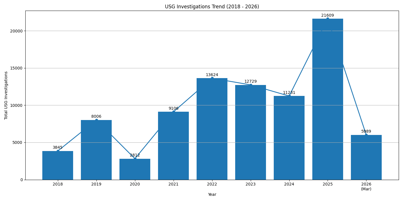
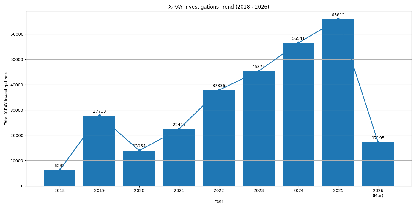
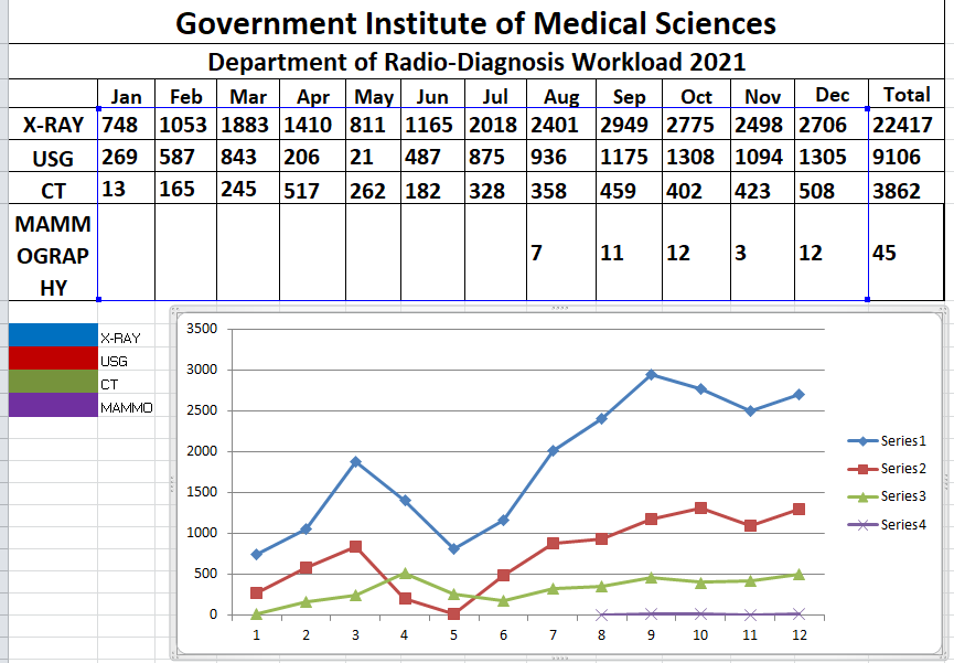
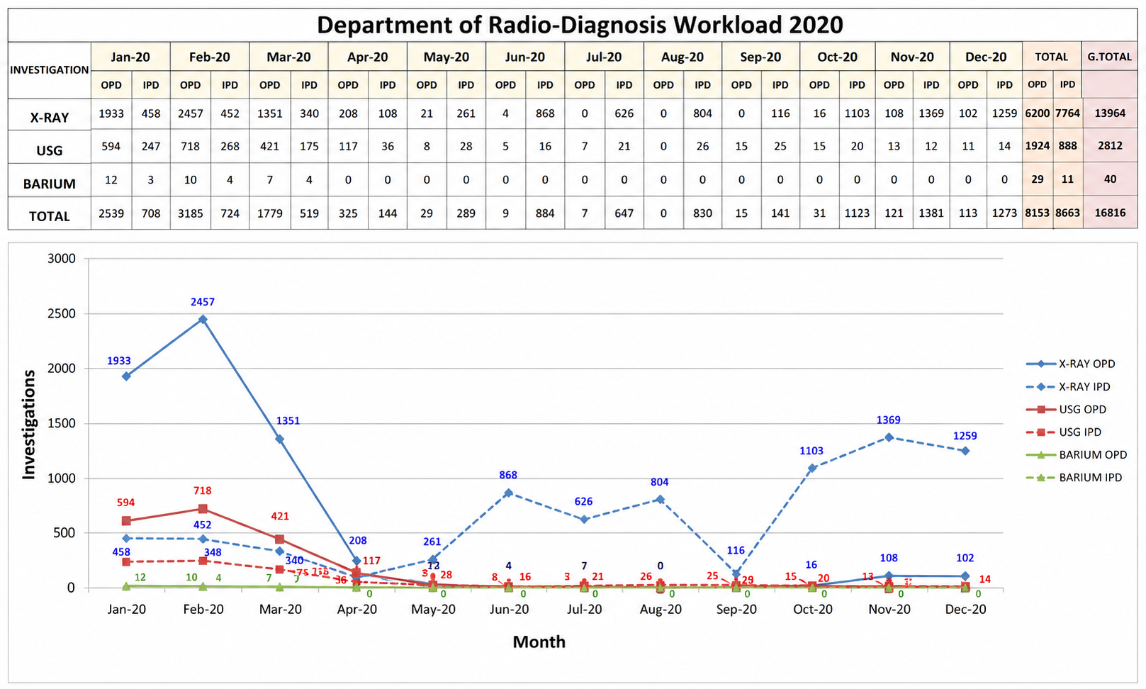
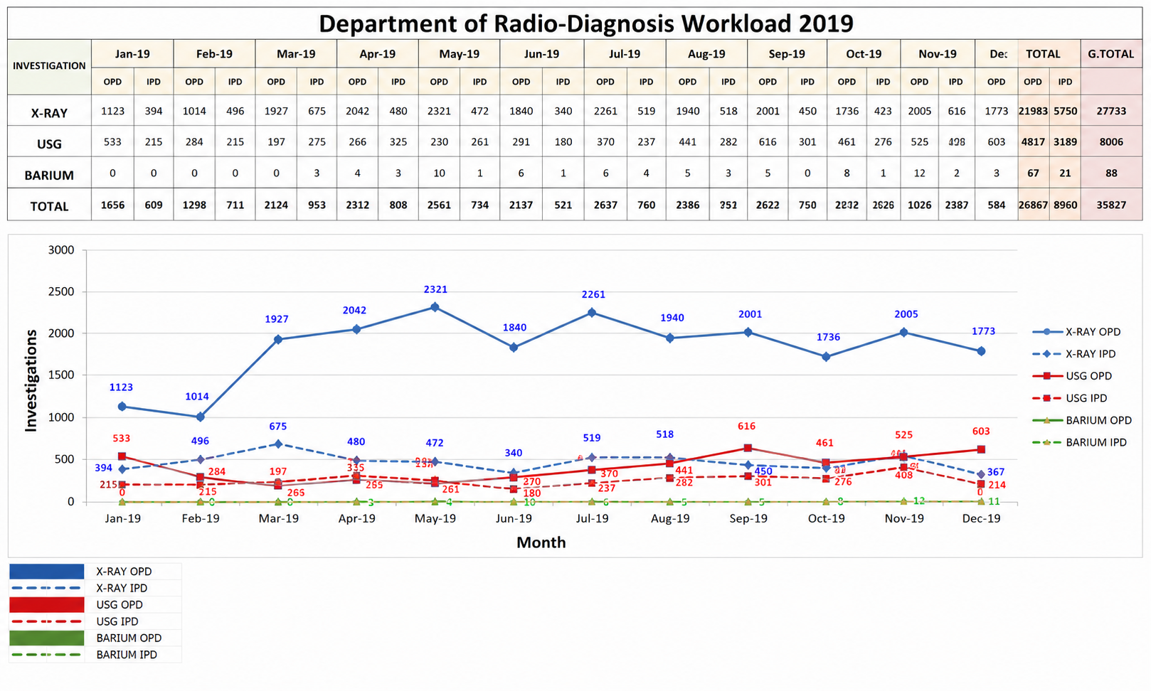
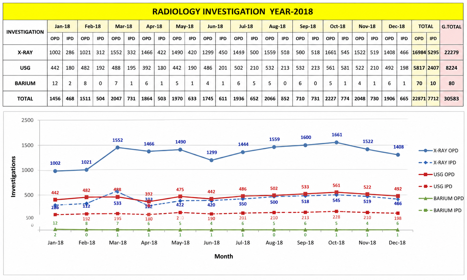
Year-wise investigation data from 2017 to March 2026. Click any card to view full screen.
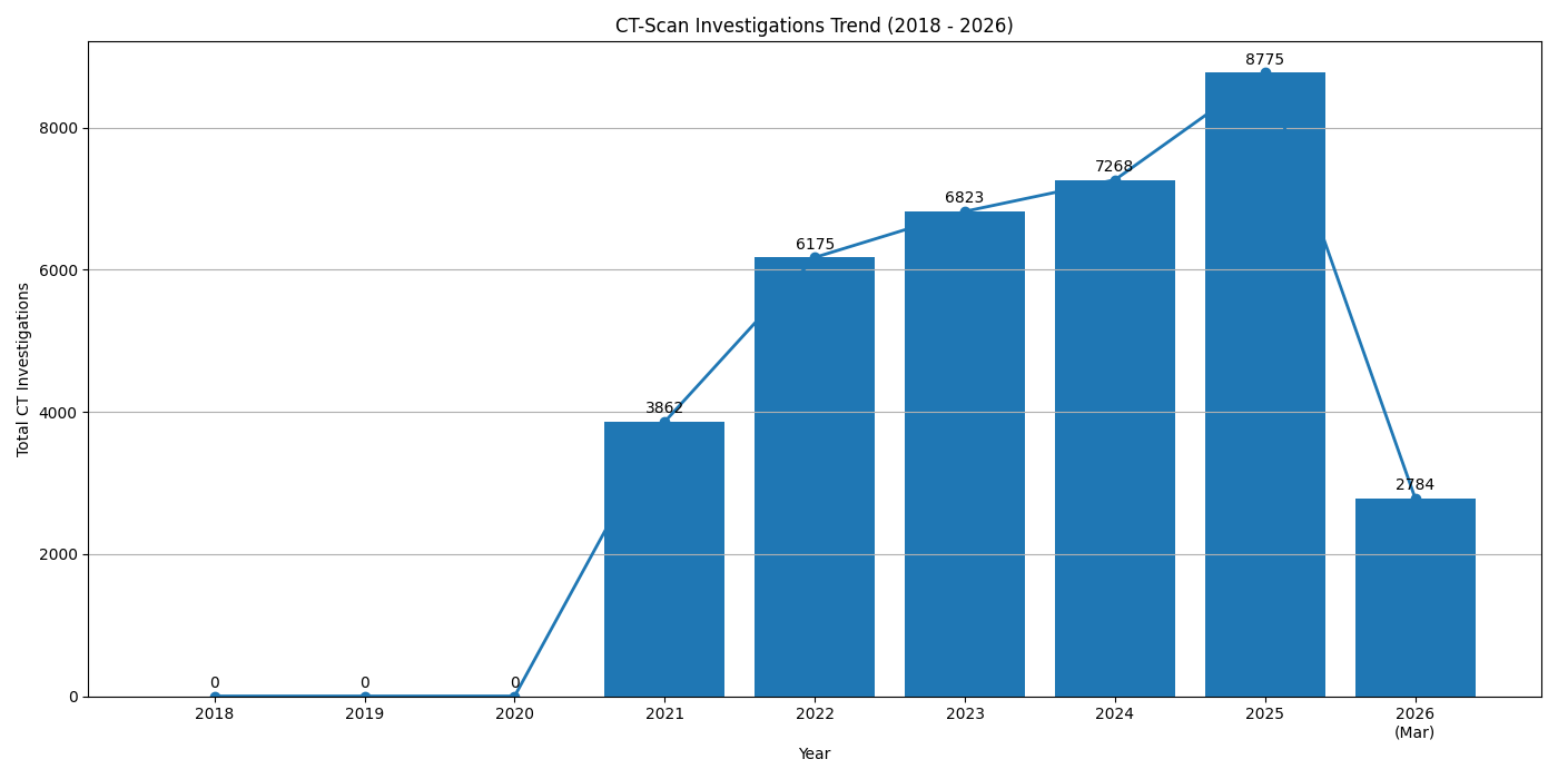
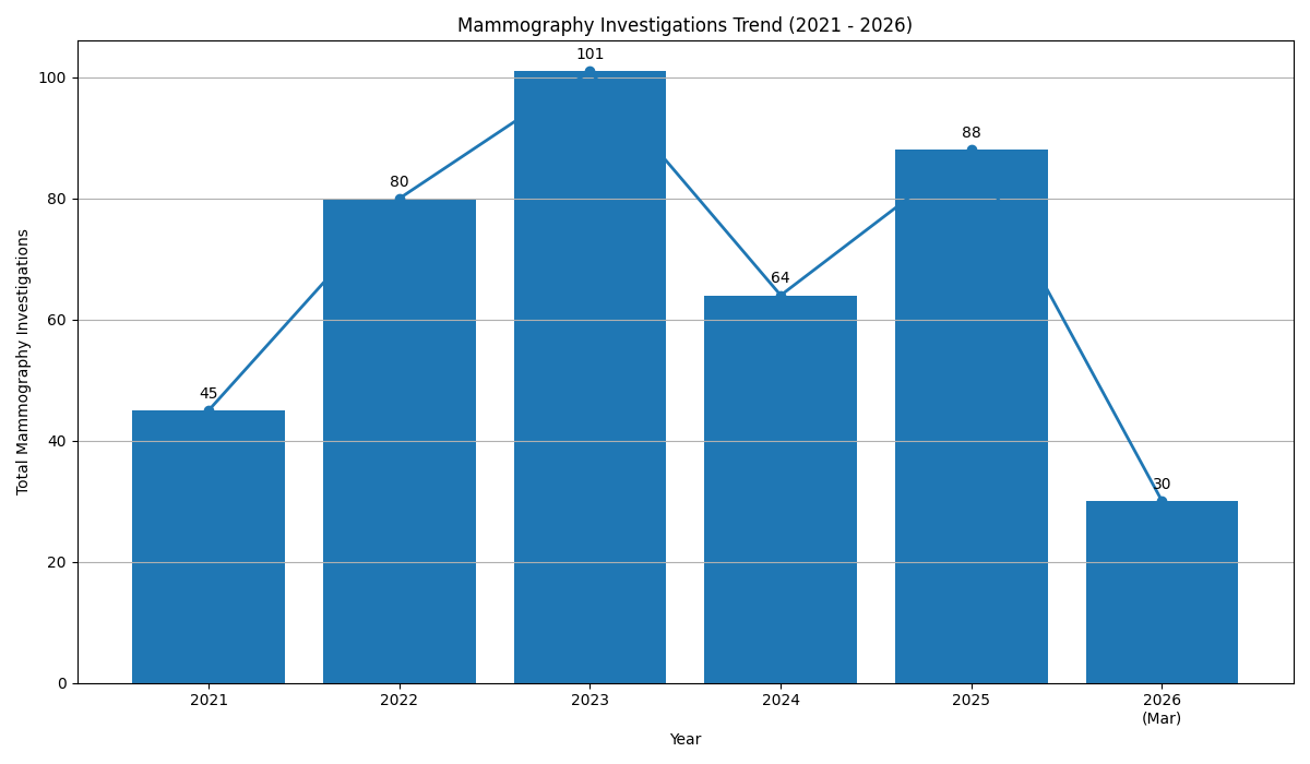
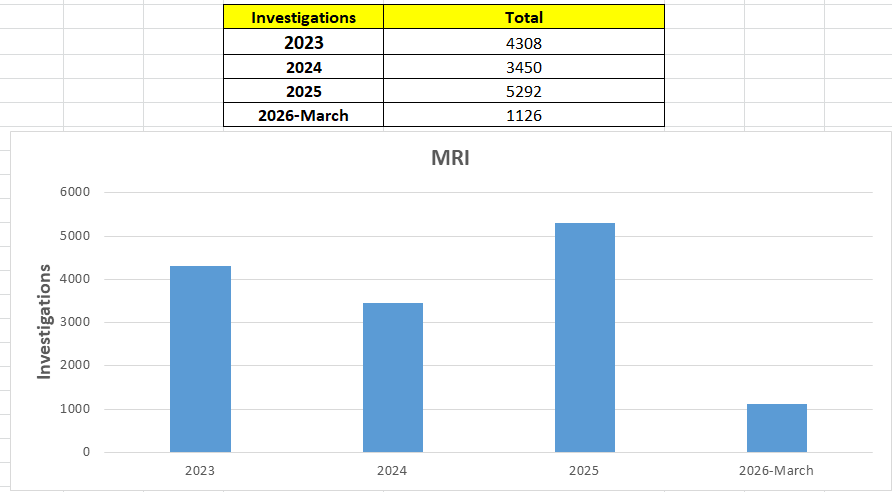
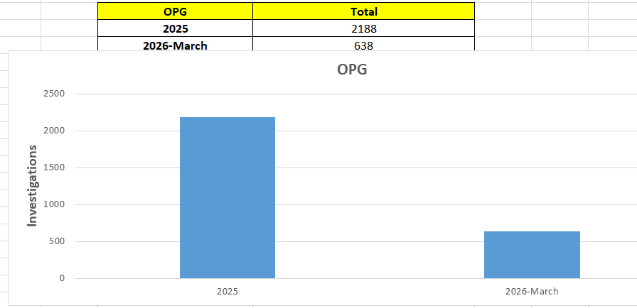
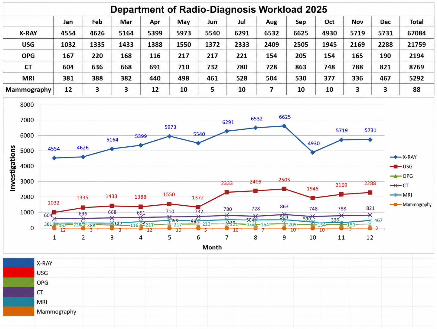
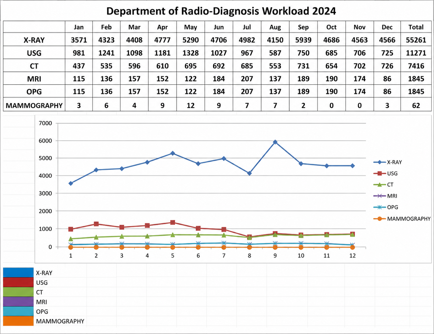
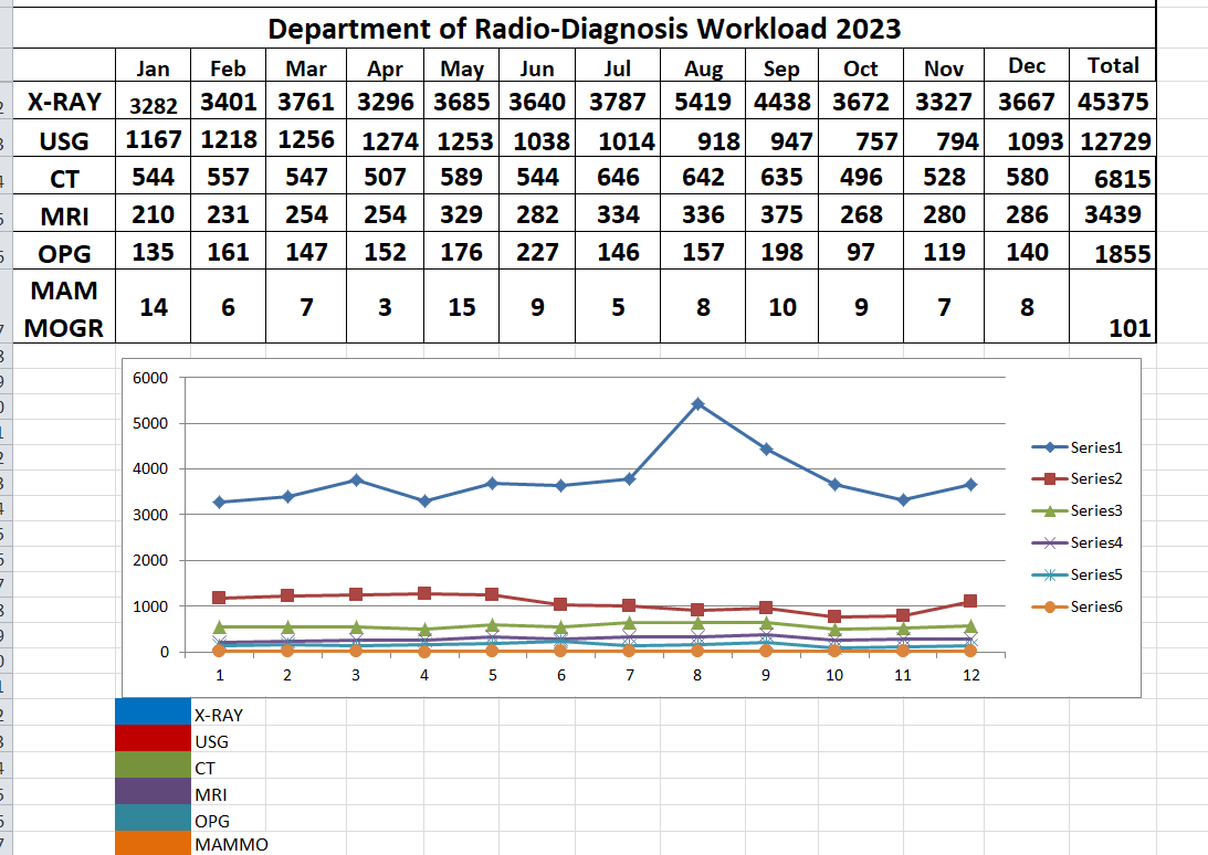
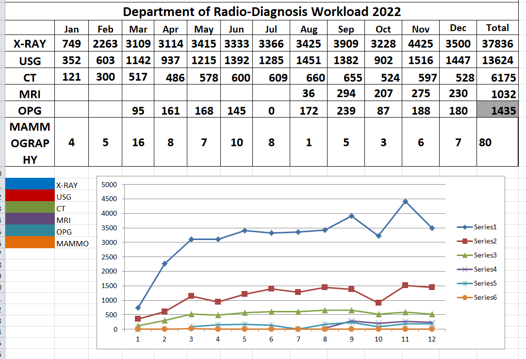
Departmental workload statistics and graphical data till 2025. Click any card to view full screen.
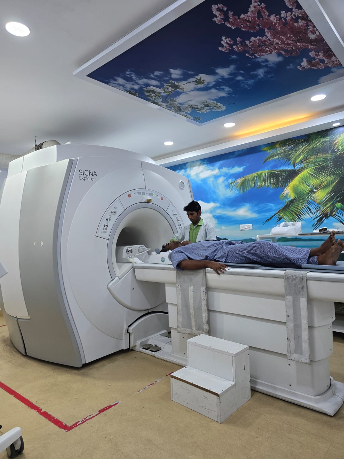
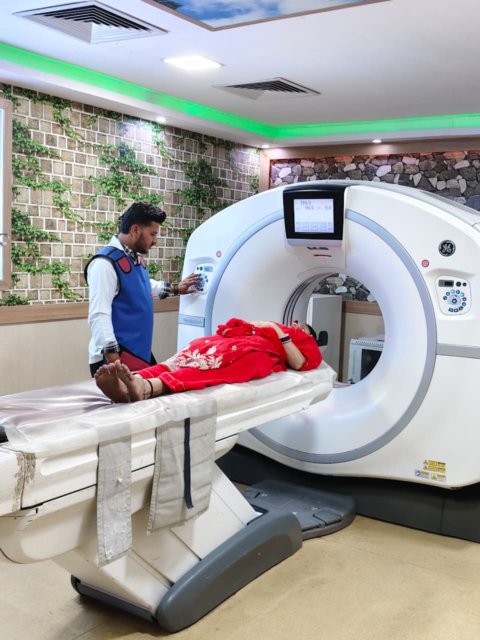
- CT Scans: 30 - 40 per Day
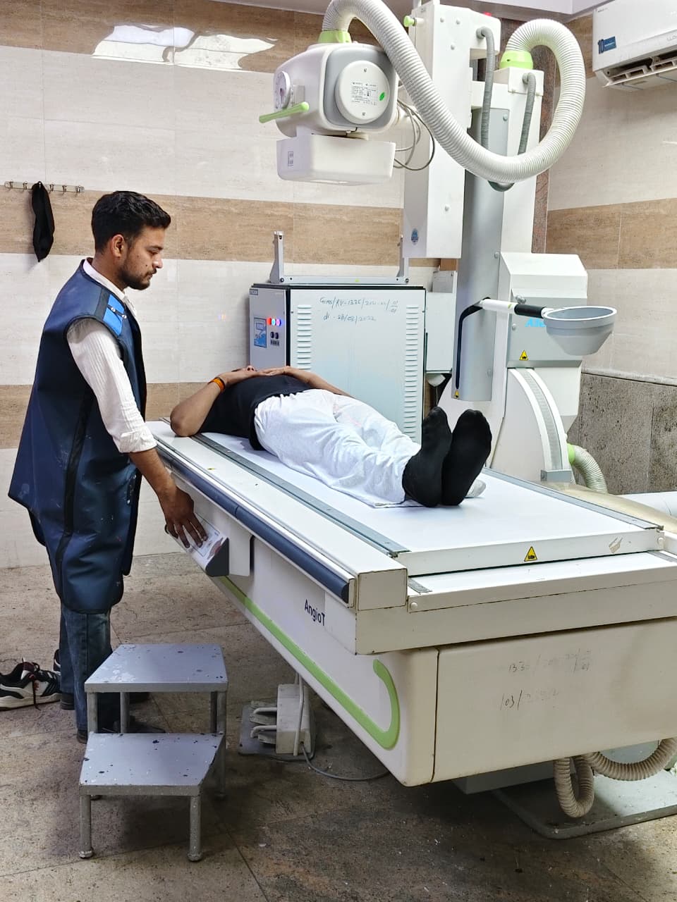
- Fluoroscopy Procedures: 2 - 3 per Day
- Interventional Procedures: 1 - 2 per Day
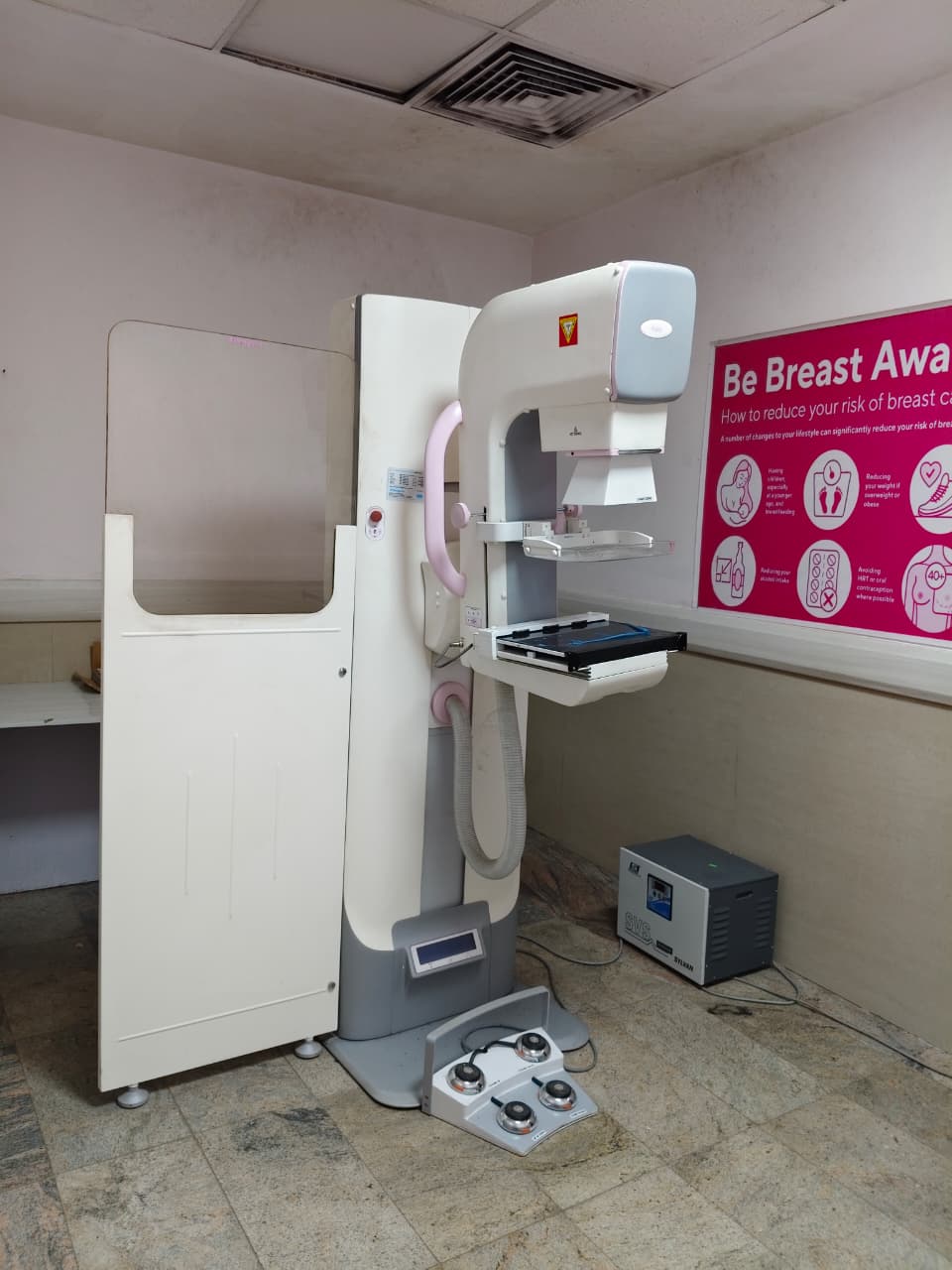
- Mammography: 1 - 2 per Day
- MRI: 10 - 15 per Day
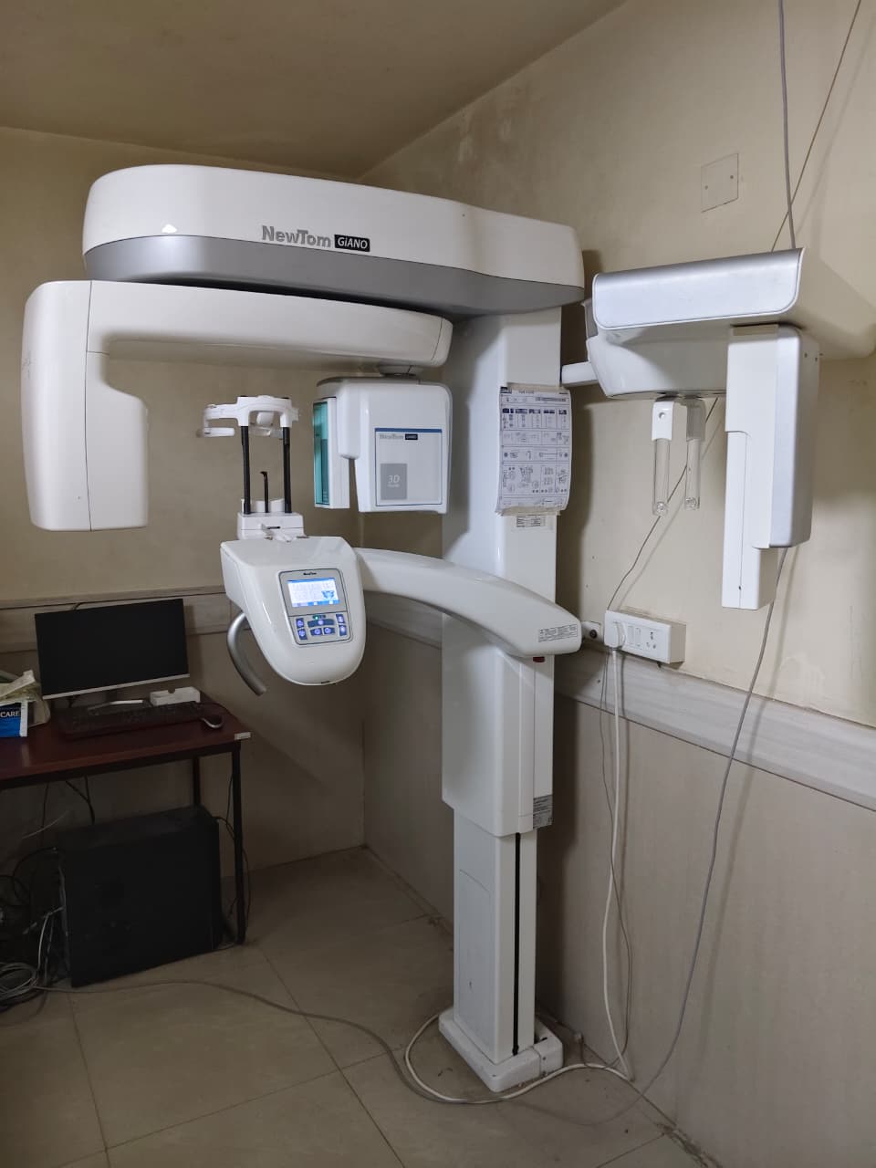
- OPG: 5 - 7 per Day
- Procedures: 3 - 4 per Day
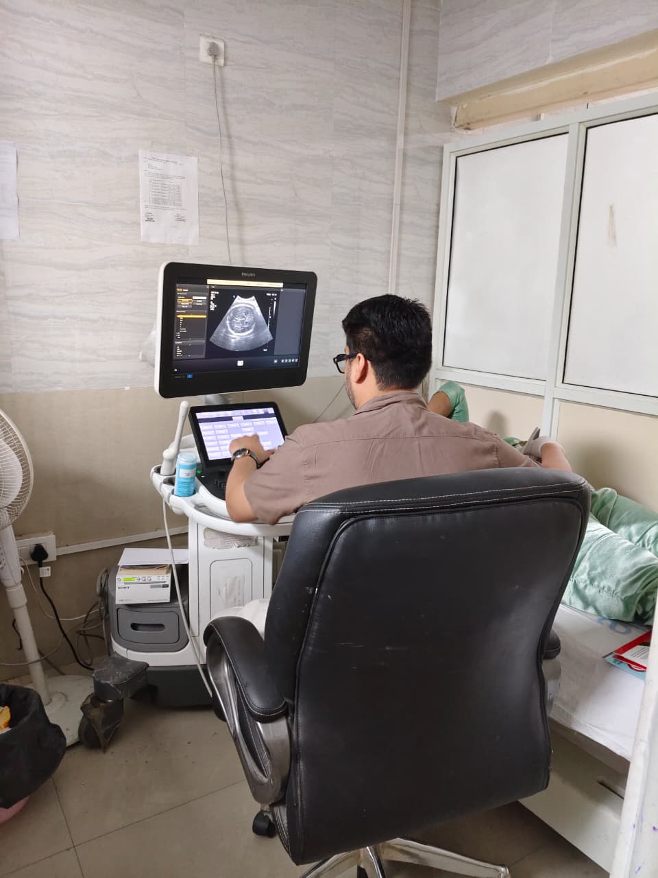
- USG: 90 - 110 per Day
- X-Rays: 150 - 200 per Day
- MBBS Programme
- DNB (Radio-Diagnosis)
- Bachelor of Medical and Radiological Imaging Technology (BMRT)
- Diploma in X-Ray Technician
- Level 1 Ultrasound Course
Duration: As per NMC norms
Authorization:
Letter of Permission granted by the Medical Council of India (MCI) vide letter No. MCI-34(41)(E-60)/2019-Med/115574 dated 20th May 2019.
MBBS course commenced w.e.f. 01st August 2019.
Duration: 3 Years
Authorization:
Accredited by the National Board of Examinations in Medical Sciences (NBEMS) vide Ref. No. NBEMS/ACCR/Granted/4318040222/418-F/2022 dated 26th September 2022.
Approved for the period January 2023 to December 2027.
Duration: 3 Years + 1 Year Internship
Authorization:
Approved by Atal Bihari Vajpayee Medical University, Uttar Pradesh and the State Government, as applicable.
Authorization:
Letter of permission from State Medical Faculty vide Ref. No. 835/71-4-2021-N-8/2016 dated 18/10/2021 vide letter No. 8558/21 dated 21/10/2021.
Eligibility: MBBS Doctors
Number of Seats: 4
Duration: 6 Months
Authorization:
Permission granted by the Government of Uttar Pradesh vide letter No. 5-8010/2023/97-2022-8 dated 23rd January 2023.
- 16th February 2020 at GIMS Greater Noida for various stakeholders in healthcare.
- Organized Seminar on Medical Ethics on 7th August 2021, at GIMS Greater Noida
- Organized a 2 day CME on 8-9th April 2022 at GIMS Greater Noida on ‘Musculoskeletal Imaging- Current Trends and Applications’ (awarded 6 credit hours from UP Medical Council)
- Organized a training on ‘Basics of Contrast Media Adverse Drug Reactions and their Management’, ‘MRI contrast safe use’ and ‘Contrast induced nephropathy prevalence & Management’ in collaboration with GE Healthcare on 14th March 2023 at GIMS, Greater Noida.
Departmental Activities
Academic & Student Engagement Initiatives
World Radiology Day Celebration — 13 November 2025
The Institute of Paramedical Sciences, GIMS Greater Noida celebrated World Radiology Day with great enthusiasm and academic spirit. The program highlighted the importance of radiology in modern healthcare and familiarized students with recent advancements in medical imaging.
The celebration began with a traditional Lamp Lighting Ceremony followed by the welcome of guest speaker Dr. Dolly A. Sharma (Professor & Head, Department of Radiology) by Dr. Saurabh Srivastav (CMS) and Dr. Manisha Singh (Principal, Institute of Paramedical Sciences). The welcome address was delivered by Dr. Jaya Bharti, Vice-Principal.
A guest lecture titled “Radiology Beyond Diagnosis” was delivered by Dr. Dolly A. Sharma, explaining the expanding role of radiology in patient care and medical research. Students also participated in a Nukkad Natak focusing on radiation safety and ethical practices, along with a debate competition on the theme “Radiation: A Necessary Evil.”
The program concluded with felicitation of winners of debate, quiz and poster competitions and a vote of thanks by Ms. Manvee Rai (Assistant Professor). The event reflected the department’s commitment toward academic excellence, student participation, and responsible use of radiological technologies in healthcare.
Event Gallery


- Get recognition for MD radiology seats.
- Become a nodal center for level 1 and 2 ultrasound courses conducted by UP government.
- Conduct relevant and significant research in collaboration with clinical departments with aim of improving diagnostic and therapeutic outcomes
- Establish organ specific subspecialty imaging services
- To enhance interventional radiology services.
- To establish simulation models for improving diagnostic and training facilities.
- Start tele radiology services and provide radiology consults online to under-served government hospitals and centers.
- Agarwal N, Jain P, Pathak R, Gupta R. Telemedicine in India: A tool for transforming health care in the era of COVID-19 pandemic. J Educ Health Promot. 2020;9:190-194
- Sharma R, Mishra P, Seth S, Agarwal N. OHVIRA Syndrome—Diagnostic Dilemmas and Review of Literature. J South Asian Feder Obs Gynae 2020; 12 (6):421-426.
- Singh JP, Agarwal N, Agarwal AK, Srivastav S. Mathematical Model on Two Layered Hepatic Blood Flow in Venules Due to Hepatitis A. Solid State Technology 2021;64 (2):5330-5338
- Agarwal N, Jain P, Khan TN, Gupta R. Chest radiographic findings and their correlation with disease progression in COVID-19 patients in northern India. J Family Med Prim Care. 2022 Feb;11(2):559-566
- Agarwal N, Jain P, Khan TN, Raja A. A retrospective study of association of CT severity with clinical profile and outcomes of patients with COVID-19 in the second wave. J Clin Imaging Sci. 2022 Apr 26;12:17A. Solid State Technology 2021;64 (2):5330-5338
- Agarwal N, Jain P, Khan TN, Raja A. Pulmonary cystic lesions in patients with COVID-19 -a case series. Journal of Clinical and Diagnostic Research. 2022 Jun, Vol-16(6)
- Jain P, Agarwal N, Saxena V, Srivastav S, Solanki H. Mortality in patients with Coronavirus disease 2019 (COVID-19) and its clinicoradiological and laboratory correlates: A retrospective study. J Family Med Prim Care 2022;11:6197-203.
- Singh JP, Agarwal N, Agarwal AK, Srivastav S. Detection of blood flow through artery in the presence of steno-occlusive disease post liver transplantation by modeling for theatrical and detrimental environmental changes. Journal of Indian Chemical Society. 2022(99): 100581
- Agarwal N, Jain P, Khan TN, Gupta R. Chest radiographic findings and their correlation with disease progression in COVID 19 patients in northern India. J Family Med Prim Care 2022;11:559-66
- Agarwal N, Jain P, Khan TN, Raja A. A retrospective study of association of CT severity with clinical profile and outcomes of patients with Covid-19 in the second wave. J Cli Imaging Sci 12 (17) (26th April, 2022)outh Asian Feder Obs Gynae 2020; 12 (6):421-426.
- Agarwal N, Jain P, Khan TN, Raja A. Pulmonary cystic lesions in patients with COVID-19 -a case series. Journal of Clinical and Diagnostic Research. 2022 Jun, Vol-16(6)
- Hepato-Biliary Ascariasis; A Suri, A Chaturvedi, M Bhatia et al. Ind J Radiol Imag. 2002-12:2:221-223.
- Port site implantation metastases. A Chaturvedi, C Mohan. AOJR 2004, 9(2&3) Apr-Sep, 81-83.
- Vein of Galen Malformation in a Neonate. A Chaturvedi, C Mohan, RG Holla. AOJR 2004,9(2&3) Apr-Sep, 106-108.
- Post Traumatic Aorto Caval Fistula. A Chaturvedi, C Mohan AOJR2004,9(2&3) Apr-Sep, 109-111.
- Spinal Cord Lipomas. HS Bhatoe, A Chaturvedi, Kavita Sahai. Neurosurgery Focus 2005,18(2) 1-5.
- Pan-ventricular Neuroblastoma. HS Bhatoe, A Chaturvedi, V Datta. J of Neuro- oncology 2005 74: 281-282.
- Imaging of Cochlear Implants. A Chaturvedi, C Mohan, SB Mahajan, V Kakkar. Ind J Radiol Imag 2006 16:3:385-392
- Review and current status of image guided interventional techniques in management of chronic low back pain. S Chaturvedi, A Chaturvedi, C Mohan. JIMSA 2007(Jan-Mar); 22:1: 101-106.
- Interventional management of aortic aneurysms. C Mohan, KM Rai, IK Indrajit, OP Mathew, A Chaturvedi. JIMSA 2007(Jan-Mar); 22:1: 57-61.
- Bronchial artery embolisation in life threatening hemoptysis. C Mohan, IK Indrajit, K Pathak, A Chaturvedi, GS Sabhiki. JIMSA 2007(Jan-Mar); 22:1:43-47.
- Cholangiocarcinoma in Choledochal cyst. A Chaturvedi, , JP Singh, V Rastogi. Ind J Radiol Imag 2008 18:3: 236-238.
- MRI diagnosis of Lymphomatosis of the Brachial Plexus. A Chaturvedi, JP Singh, V Rastogi. Neurology India 2008 56: 4: 97-98.
- Image Guided Lumbar Facet Joint Infiltration in Non Radicular Low Back Pain. A Chaturvedi, S Chaturvedi, R Siva Shankar. Indian Journal of Radiology & Imaging 2008 19:1: 28-34.
- Cecil DM, Chaturvedi A, Kapoor D. Z-shaped brainstem and other magnetic resonance imaging findings in congenital muscular dystrophy. Neurol India 2016;64:577 Role of MRI -8
- Teflon Granuloma: A rare cause of recurrent trigeminal neuralgia. Sharma G, Chaturvedi A, Kapoor D. Neurol India 2015,63:803-4.
- Role of MRI in post ACL reconstruction Complications. Dr Garima Sharma, Dr Arti Chaturvedi, Dr Deepti Cecil. Oral Paper presented at 71st IRIA conference 2018.
Outreach Programme
Department of Radio-Diagnosis — GIMS, Greater Noida
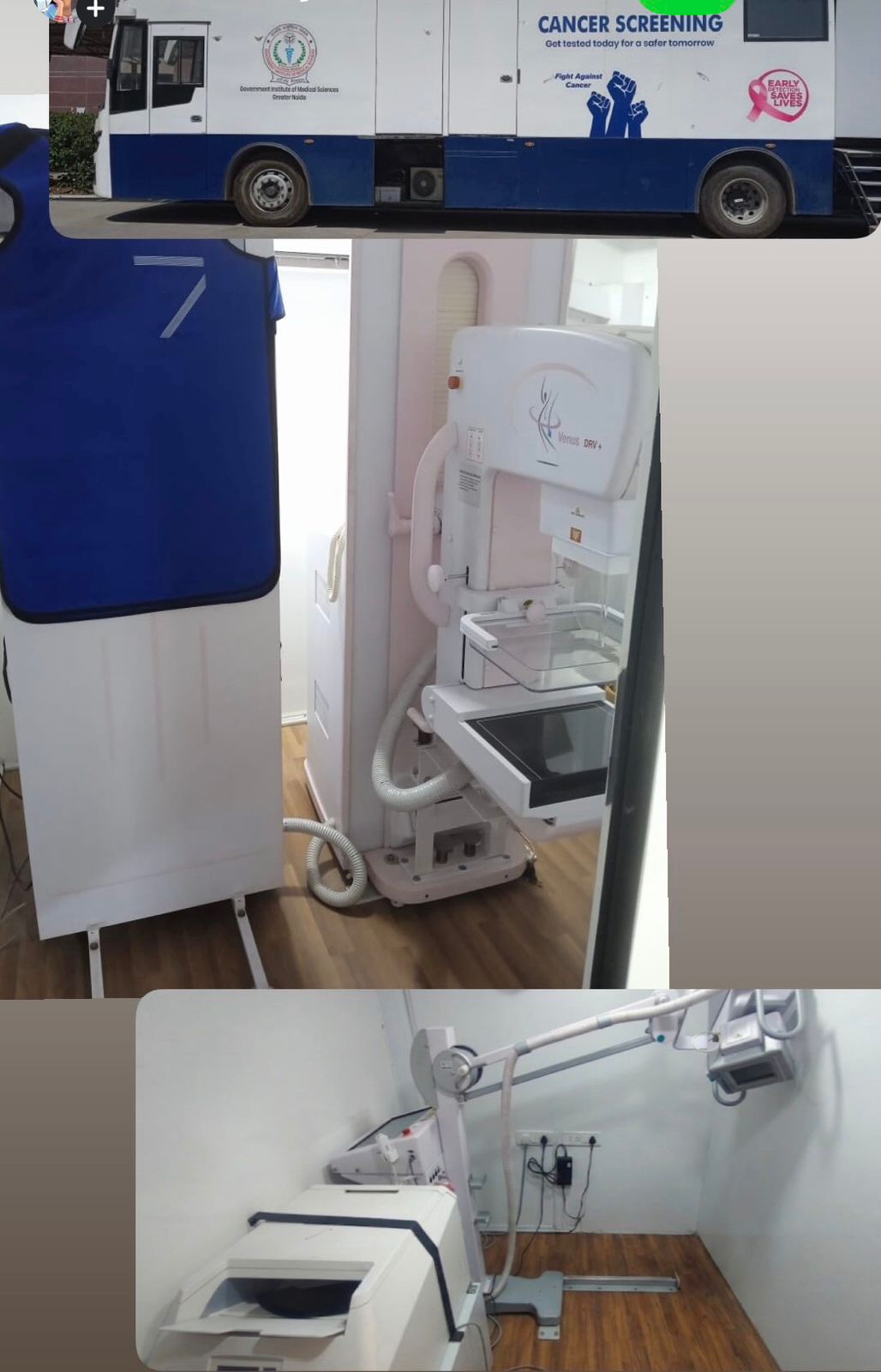
At GIMS, the Department of Radio-Diagnosis organizes a weekly schedule for departmental outreach and community visits. These initiatives are made possible through our Mobile Cancer Screening Van, which serves as a significant benefit for underprivileged communities and asymptomatic individuals by providing easy access to screening services.
Advanced PACS & AI Integrated Imaging System
Our department utilizes AI-driven image analysis to enhance diagnostic accuracy and streamline reporting workflows.
- Automated workflows for efficient case prioritization.
- Data-driven insights supporting clinical outcomes.
- Future-ready radiology infrastructure.
Mobile Cancer Detection Van Services
Delivering advanced screening services directly to the community:
- On-site Mammography: Screening for breast cancer in asymptomatic women.
- Chest X-ray: Evaluation of skeletal and thoracic disorders.
- Early Detection: Promoting preliminary diagnosis in rural and urban areas.
- Health Awareness: Dedicated education programmes for preventive care.
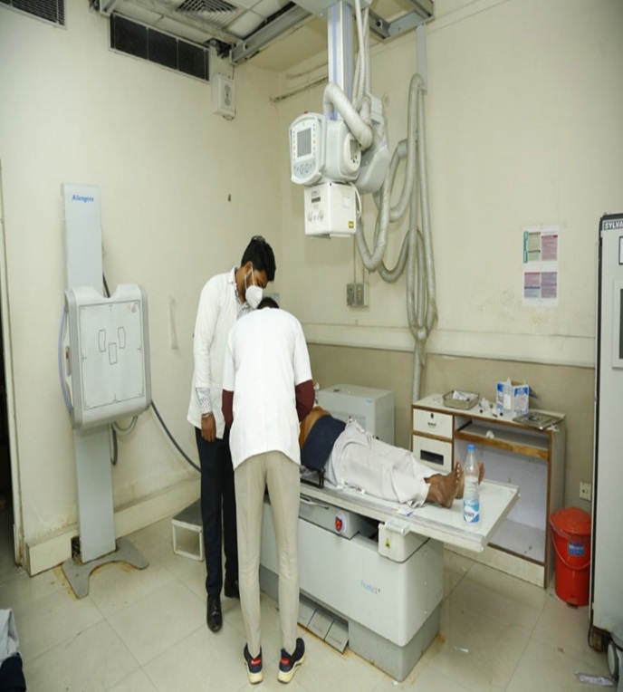
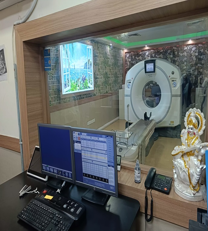
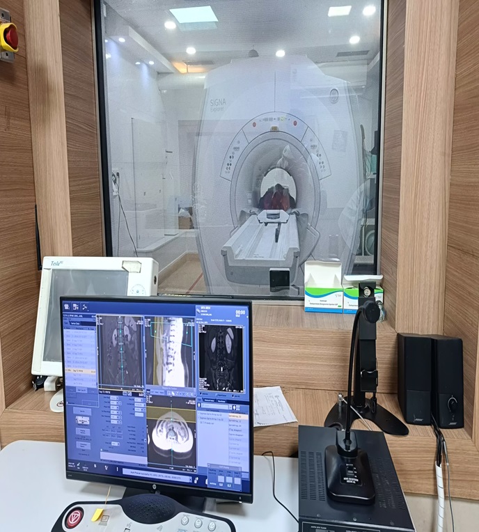
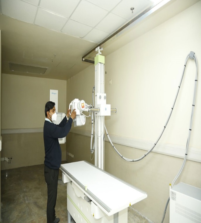
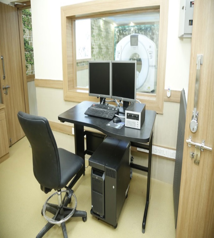
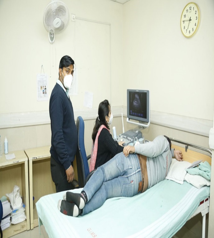
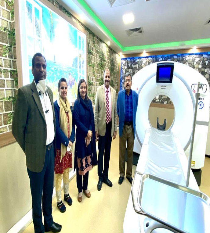
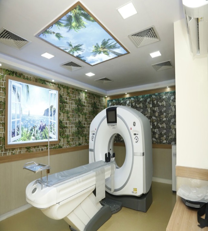
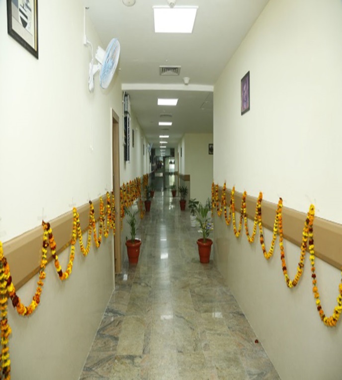
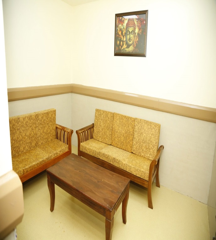
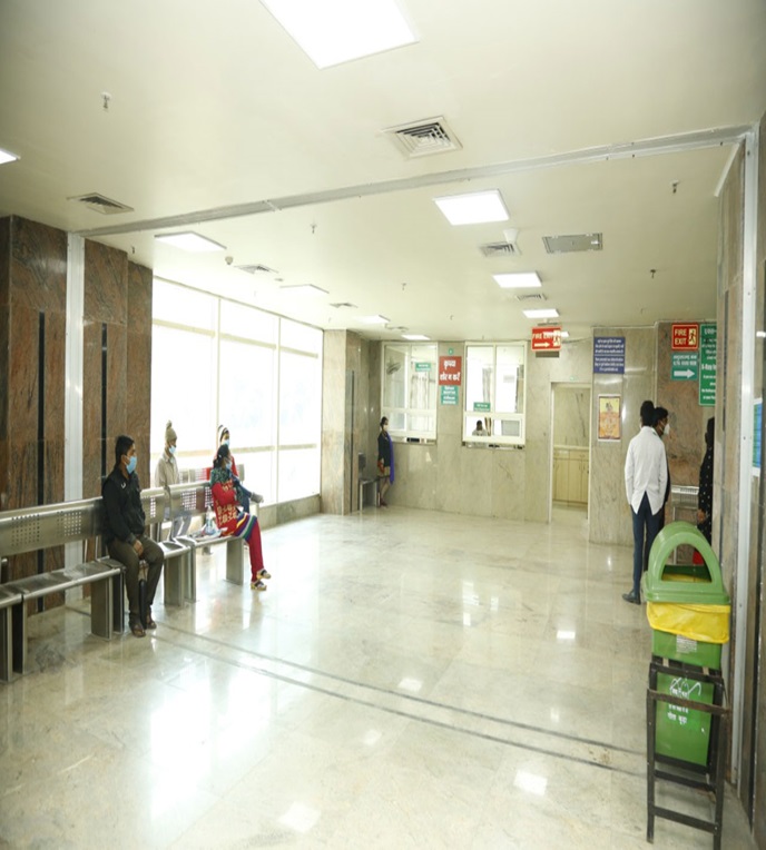
Department Gallery
DNB Monthly Teaching Schedule
Department of Radiodiagnosis – Academic Calendar
| S.NO | DAYS | TYPE OF CLASS | MODERATOR |
|---|---|---|---|
| 1 | MONDAY | Ultrasound related topic | Dr Rachna Chaurasia |
| 2 | TUESDAY | Seminar | Dr Rachna Chaurasia |
| 3 | WEDNESDAY | X-Ray trainee classes – DXRT / BMRIT | Dr Rachna Chaurasia |
| 4 | THURSDAY | Journal club | Dr Rachna Chaurasia |
| 5 | FRIDAY | Radio Physics | Dr Rachna Chaurasia |
| 6 | SATURDAY | Case / Tutorial | Dr Rachna Chaurasia |
Post-Graduate (DNB) Examination Results
Academic Session — University Examination Outcome